
Figure 6.
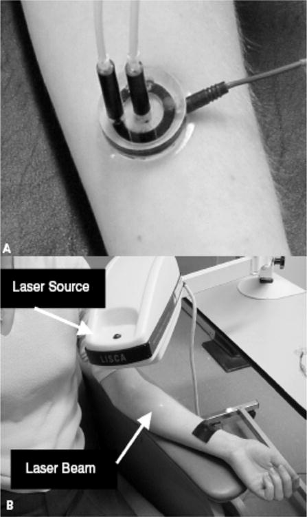
Measurement of skin microcirculation. (a) A small quantity of (<1ml) of 1% Ach chloride solution or 1% sodium nitroprusside solution is placed in the iontophoresis chamber. A constant current of 200 mA is applied for 60 seconds achieving a dose of 6 mC/cm2 between the iontophoresis chamber and a second nonactive electrode placed 10 to 15 cm proximal to the chamber (black strap around the wrist). This current causes a movement of solution to be delivered toward the skin. (b) Laser Doppler flowmetry: A helium-neon laser beam is emitted from the laser source to sequentially scan the circular hyperemic area produced by the iontophoresed vasoactive substance to a small area on the volar surface of the forearm. Figure adapted with permission from A. Veves (145).