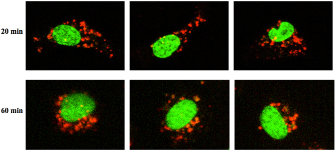
Figure 3.

Live confocal microscopy visualizing STBEV uptake and intracellular transport. Uptake of PKH-labelled STBEVs into HCAECs was followed by live confocal microscopy. Three representative cells are shown after 20 and 60 min of incubation. After 20 min the STBEVs are clearly internalised and widely distributed in the cell cytoplasm. After 60 min the STBEVs appear to be closer to the cell nuclei. Cell nuclei are pseudocolored in green, and PKH-labelled STBEVs in red. The figures are from live imaging, explaining the lower intensity at 60 minutes due to bleaching.
