
Figure 6.
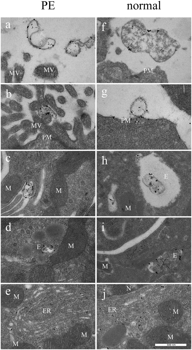
STBEV uptake and miRNA transfer visualised by transmission electron microscopy. By using TEM, PE STBEVs (a–e) and normal STBEVs (f–j) were visualized in HCAECs. In panel (a), PE STBEVs approach the HCAEC. In (b) and (c), PE STBEV appears to be binding to the ruffled plasma membrane. The STBEV is seen inside the cells in what appears to be an endosome (d), in close proximity to the mitochondria, endoplasmic reticulum and cell nucleus. The mir-517c from PE STBEVs appears to be in larger number in the ER (e) compared to the mitochondria. PLAP appears to stay in the endosomes and is possibly recycled to the cell membrane. The normal STBEVs, which also carry mir-517c are found outside the cells (f–g) and can be found in endosomes (h,j). In contrast to PE STBEVs, mir-517c from normal STBEVs are localized in a higher degree to mitochondria and in lower quantity to the ER (j). The STBEV and miRNA intracellular distribution are also described in Table 1. Abbreviations; E endosome, ER endoplasmic reticulum, MV microvilli, M mitochondria, N nucleus, PM plasma membrane. PLAP labelled with 20 nm and mir-517c labelled with 5 nm colloidal gold.