
Figure 7.
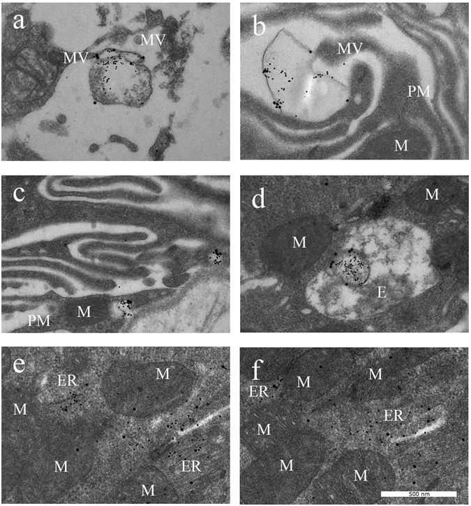
STBEV uptake and effect of free HbF visualised by transmission electron microscopy. The normal STBEVs, co-treated with HbF, appear to bind the plasma membrane (a), which is ruffled (b,c) as was also seen in cells treated with PE STBEVs (see Fig. 6b). The STBEVs were internalised into endosomes (d) and transported to intracellular compartments (e,f). The mir-517c is localised predominantly into the ER than in the mitochondria (e,f), also seen in cells treated with PE STBEVs (see Fig. 6e). Abbreviations; E endosome, ER endoplasmic reticulum, MV microvilli, M mitochondria, N nucleus, PM plasma membrane. PLAP labelled with 20 nm and mir-517c labelled with 5 nm colloidal gold.