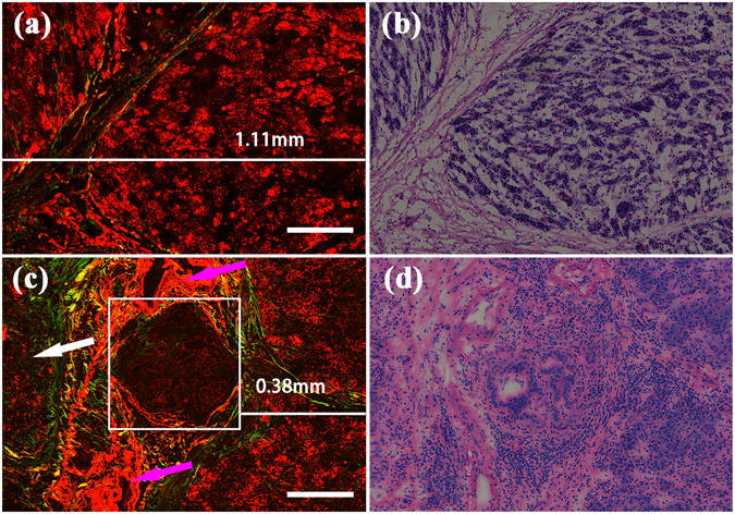
Figure 7.

MPM images and corresponding H&E-stained images of R0 and R1 resections of the pancreatic neck margin. Magnification of H&E-stained images is 40 ×; Scale: 200 μm. (a) MPM image of R0 resection; (b) The corresponding H&E-stained image of R0 resection; (c) MPM image of R1 resection; (d) The corresponding H&E-stained image of R1 resection. White arrows: increased collagen fibres; pink arrows: blood vessels; white box: neoplastic glands.
