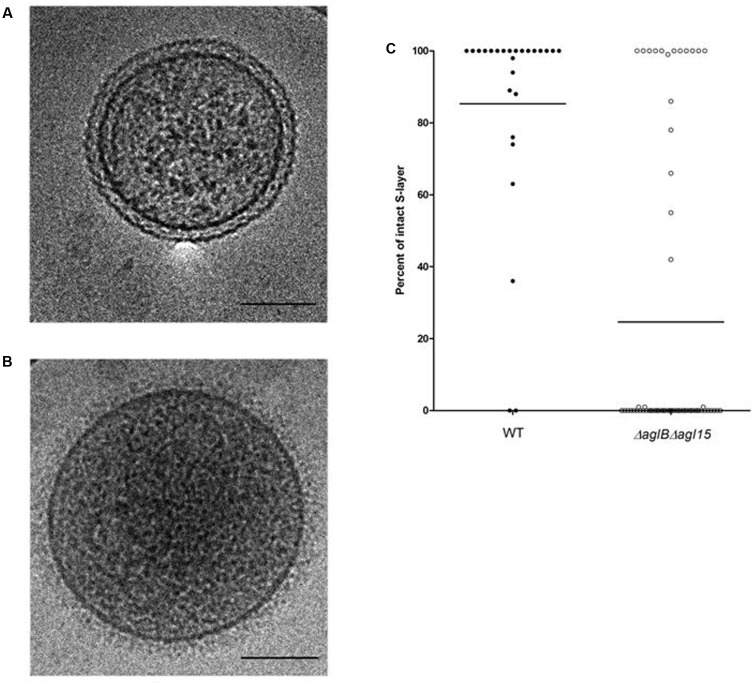
FIGURE 6.
Cryo-transmission electron microscopy of right-side-out membrane vesicles. The S-layer is compromised in RSO membrane vesicles prepared from H. volcanii ΔaglBΔagl15. (A,B) Electron micrograph of a representative RSO membrane vesicles derived from H. volcanii cells of the WT glycosylation strain (A) and the ΔaglBΔagl15 strain (B). In each panel, the bar represents 100 nm. (C) The percentages of intact S-layer in RSO membrane vesicles prepared from cells of the WT glycosylation strain (left, full symbols; n = 26) and the ΔaglBΔagl15 strain (right, open symbols; n = 62). In each column, the average (horizontal line) and the distribution of values collected from each vesicle population are presented.

