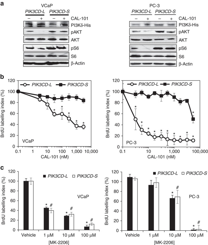
Figure 5. PI3Kδ-S but not PI3Kδ-L is resistant to small-molecule inhibition of PI3K/AKT/mTOR signalling and proliferation.
(a) Assessment of PI3K/AKT/mTOR signalling following treatment with vehicle (saline) or CAL-101 (100 nM, 24 h). PI3K/AKT/mTOR signalling was assessed by western blot analysis with phospho-antibodies to AKT (pAKT) and S6 ribosomal protein (pS6). β-Actin served as a loading control. His-tag antibody was used to demonstrate equal expression of His-tagged variant PI3Kδ protein in stably transfected cell lines. Representative images from at least three independent western blot experiments. Unprocessed western images shown in Supplementary Fig. 12. (b) Proliferation in VCaP and PC-3 cells stably overexpressing the PIK3CD-S variant or PIK3CD-L variant following treatment with vehicle (saline) or selective PI3Kδ small molecule inhibitor CAL-101 (24 h). Data presented as mean±s.e.m. from at least four independent experiments for each treatment group. *Significantly different from -S variant, P<0.05 by ANOVA with Dunnett’s post hoc test. (c) Treatment of PIK3CD variant-overexpressing cells with vehicle (saline) or selective AKT small molecule inhibitor MK-2206 (24 h). Proliferation was assessed using a 5-bromodeoxyuridine (BrdU) labelling assay. Data presented as mean±s.e.m. from at least four independent experiments for each treatment group. * Or #significantly different from corresponding vehicle control, P<0.05 by ANOVA with Dunnett’s post hoc test. Variance was similar among groups being compared.

