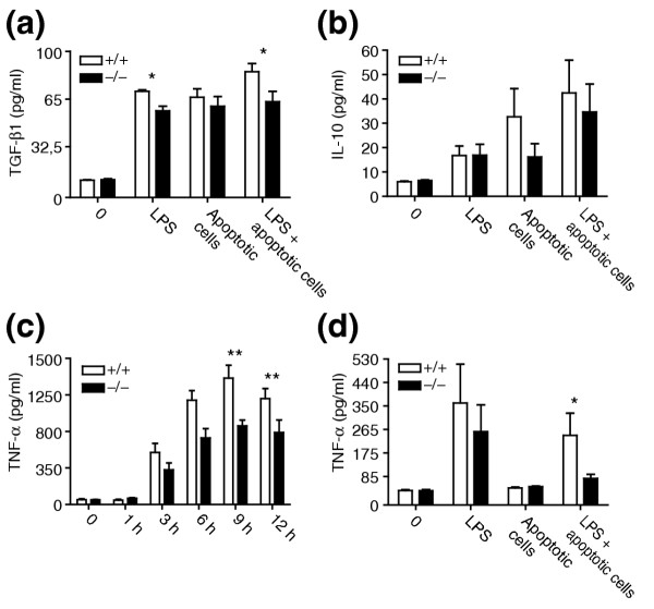
Figure 8.

Cytokine production by FLDMs upon stimulation with lipopolysaccharide (LPS) and apoptotic cells. FLDMs from wild-type and Ptdsr -/- embryos were incubated (a,b,d) with medium (0), LPS (10 ng/ml), apoptotic cells (ratio 1:10) or in combination with LPS and apoptotic cells or (c) with LPS (100 ng/ml) alone. Culture supernatants were harvested after 22 h (a,b,d) or at the indicated time points (c). TNF-α and TGF-β1 were quantified by ELISA and IL-10 by cytometric bead array (CBA) assay. Data are presented as mean ± SEM from at least three independent experiments, each carried out in triplicate. *, significant difference between genotypes, p < 0.05; **, significant difference between genotypes, p < 0.01; Wilcoxon-signed rank test.
