1.
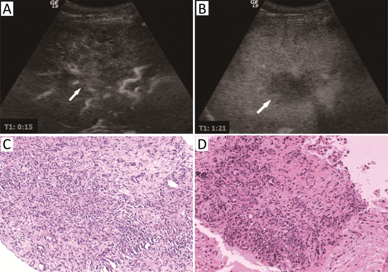
Contrast-enhanced ultrasound (CEUS) images and pathological findings in a 32-year-old female patient who suffered from fever leading to the finding of a space-occupying lesion in the liver. (A) Arterial phase showed flaky hypo-enhancement; (B) The lesion washed out at 1 min 21 s. The inflammatory lesion was suspected after CEUS examination; (C) Pathological result of the first liver biopsy showed a large number of inflammatory cell infiltration; (D) Pathological result of the second liver biopsy carried out one month later showed a small number of malignant cells among a large quantity of fibrous tissue. The patient was eventually diagnosed as intrahepatic cholangiocarcinoma (ICC).

