2.
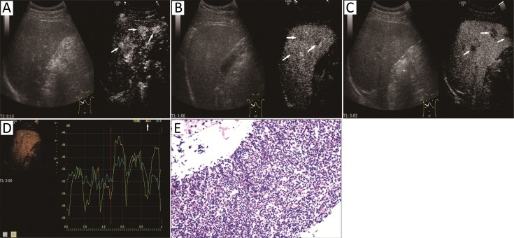
A 42-year-old male with multiple liver lesions found on routine examination which was misdiagnosed as malignant on contrast-enhanced ultrasonography (CEUS). (A) Multiple hyper-enhanced liver lesions with unclear border were found during arterial phase on CEUS (white arrows); (B) The starting washout time was 66 s; (C) The lesions washed out obviously on 3 min; (D) On time-intensity curve (TIC) analysis, the intensity of the lesion and liver parenchyma was –54.4 (yellow) and –51.5 (blue) respectively. Based on CEUS performance, the liver lesions were suspected as malignant; (E) Microscope after liver biopsy showed a large number of eosinophils cell diffuse infiltration and some small abscesses formation. The pathology suggested the possibility of parasitic infection.

