
Extended Data Figure 3.
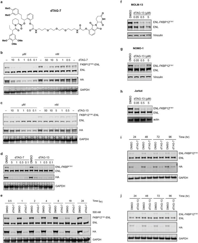
(a) Chemical structure of dTAG-7.
(b) Dose-responsive FKBP12F36V-ENL degradation detected by immunoblot after 16 hour treatment of MV4;11 (Cas9+, FKBP12F36V-HA-ENL+) cells with DMSO (labelled with “− “) or dTAG-7 at the indicated concentration.
(c) As in (b), but with dTAG-13.
(d) Immunoblot detection of ENL-FKBP12F36V degradation in MV4;11 (Cas9+, ENL-FKBP12F36V-HA+) after 16 hours.
(e) Kinetic evaluation of FKBP12F36V-ENL degradation by dTAG-7 (500 nM) in MV4;11 (Cas9+, FKBP12F36V-HA-ENL+) cells.
(f) Degradation of ENL-FKBP12F36V in MOLM-13 cells following a 4-hour treatment of ENL-FKBP12F36V-expressing cells with dTAG-13 (500 nM).
(g) As in (f), but with NOMO-1 cells expressing ENL-FKBP12F36V.
(h) As in (f), but with JURKAT cells expressing ENL-FKBP12F36V.
(i) Long-term kinetic evaluation of ENL-FKBP12F36V degradation by a single dose of dTAG-7 (500 nM) in MV4;11 (Cas9+, ENL-FKBP12F36V-HA+) cells.
(j) As in (i), but with dTAG-13 (500 nM).