
Figure 2.
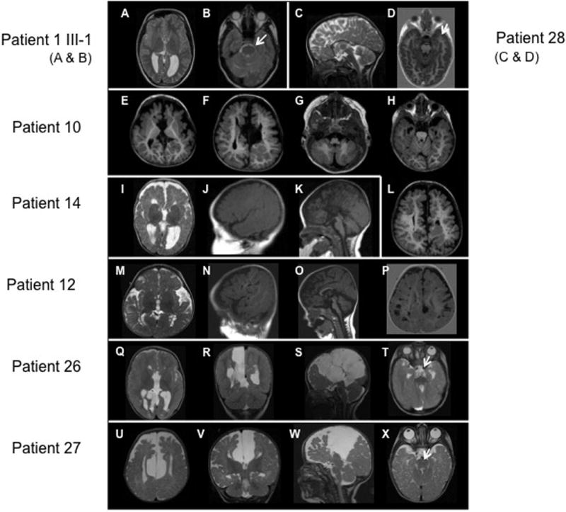
Brain MRIs of patients with OFD1 mutations. A, B: Patient II-1 in family 1, showing mild asymmetry with the right frontal lobe smaller than the left (A, B), reduced white matter volume, and mildly enlarged lateral ventricle (A). C, D: Patient 28 showing a short, thin, and dysmorphic corpus callosum. E–H and L: Patient II-2 in family 10, showing a large interhemispheric cyst (F), I–K: Patient 14, showing reduced white matter volume (I), mildly enlarged and widely separated lateral ventricles (I), severe partial agenesis of the corpus callosum (K), and possibly subtle cerebellar hypoplasia (K). M–P: Patient 12, showing a prominent bossed forehead (N, O), numerous large perivascular (Virchow–Robin) spaces in the white matter (mostly posteriorly) (N and P), a short, thin corpus callosum, and mild cerebellar vermis hypoplasia (O). Q–T: Patient 26, showing multiple, large interhemispheric cysts displacing the right hemisphere laterally (Q–S), reduced white matter volume (Q, R), a markedly enlarged third ventricle with an uncertain connection to the interhemispheric cysts (Q, R), and complete agenesis of the corpus callosum (Q, S). U–X: Patient II-1 in family 27, showing a large interhemispheric cyst displacing both hemispheres laterally (U–W), reduced white matter volume, particularly frontally (U, V), a markedly enlarged third ventricle that appears to connect to the interhemispheric cysts (V, W), complete agenesis of the corpus callosum (W), mildly small cerebellum with abnormally small anterior vermis, and mildly small posterior fossa (W).