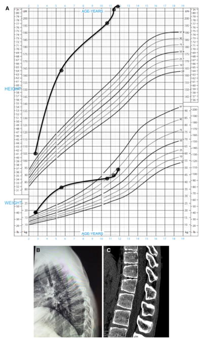
Fig. 1.
a Growth chart of the patient, with excess height and weight already being established at the age of 2 years 7 months. The patient’s height has remained excessive throughout this period, but his weight fell back into the normal range (90th percentile) due to poor nutrition, which has been addressed. b Lateral radiograph of the thoracic spine illustrating kyphosis. c Helical CT scan of the thoraco-lumbar region illustrating irregularities of the vertebral plateau and an intervertebral vacuum phenomenon (circled). Such vacuum phenomena are due to gas (nitrogen) accumulation from sources such as Schmorl node formation in juvenile kyphosis or osteoarthritis [19]

