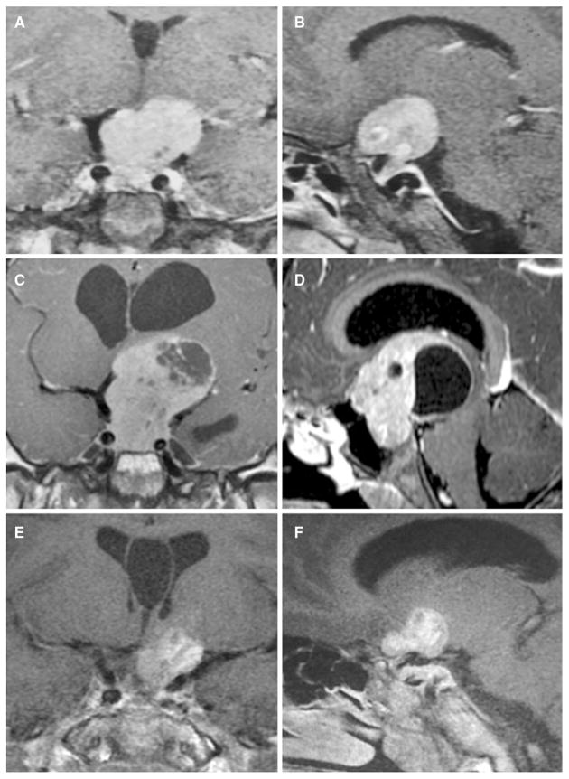
Fig. 2.
a, b Coronal and sagittal T1-weighted MRI taken at the first presentation (age of 5 years and 8 months) images showing large homogeneously enhanced pituitary macroadenoma, without cavernous sinus invasion or encasement of internal carotids. Coronal and sagittal T1-weighted MRI taken at age of 10 years. c, d Showing a marked increase in the size of the pituitary mass. The top of the tumor reaches the floor of the lateral ventricles, which are dilated. A heterogeneous region of necrotic/degenerative change is seen principally in the upper part of the tumor. Coronal and sagittal T1-weighted MRI with contrast taken three months after surgical debulking. e, f Showing pituitary macroadenoma remnant with cystic degeneration and resolution of the hydrocephalus. An incidental cyst of the septum pellucidum is seen on multiple images

