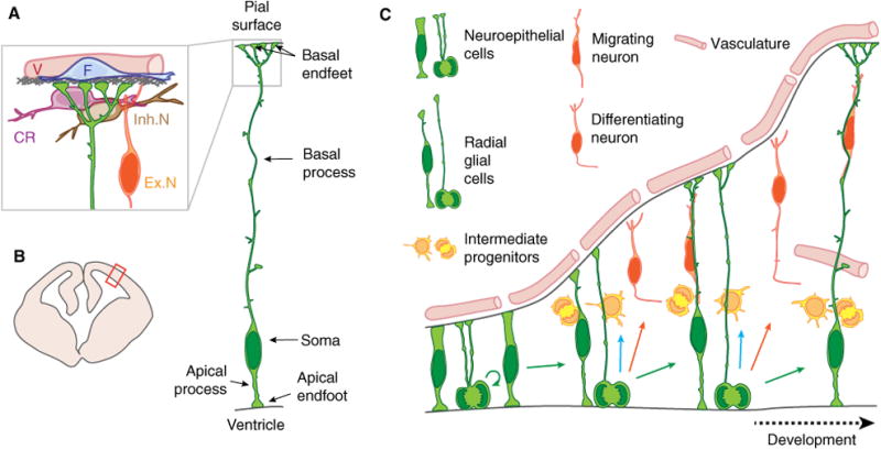
Fig. 1.

Cartoon of developing brain and neural stem cells with highlighted anatomy of a radial glial progenitor cell. (A) Anatomy of a radial glial progenitor and the endfoot niche, including the basement membrane (gray), inhibitory neurons (Inh.N), excitatory neurons (Ex.N), cajal retzius cells (CR), vasculature (V), and fibroblasts (F). (B) Schematic representation of a coronal section of an embryonic mouse brain during midcorticogenesis. Red box points to the location represented in (A). (C) Cartoon representation of mouse cortical development. This panel shows the different cell types referred to in the present paper. During early corticogenesis, neuroepithelial cells divide symmetrically to expand the precursor pool. As development proceeds, neuroepithelial cells convert into radial glial cells that mainly divide asymmetrically to produce a new RGC and either a neuron or an IPs. IPs divide away from the ventricular border to generate neurons.
