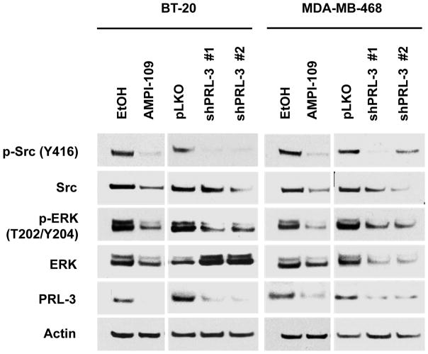
Figure 1. AMPI-109 treatment and PRL-3 knock down inactivate Src, ERK signaling in TNBC cells.
Immunoblot analysis of changes in activation of Src and ERK proteins as assessed by phosphorylation of Src at tyrosine 416 (Y416) and ERK at threonine 202 (T202) and tyrosine 204 (Y204) in vehicle control (EtOH) or AMPI-109 (100 nM) treated BT-20 and MDA-MB-468 TNBC cells. Right panels for each cell line depict changes in Src and ERK activation in PRL-3 knock down (shPRL-3 #1 and shPRL-3 #2) compared to non-silencing control cells (pLKO). The experiment was repeated twice and representative images shown.

