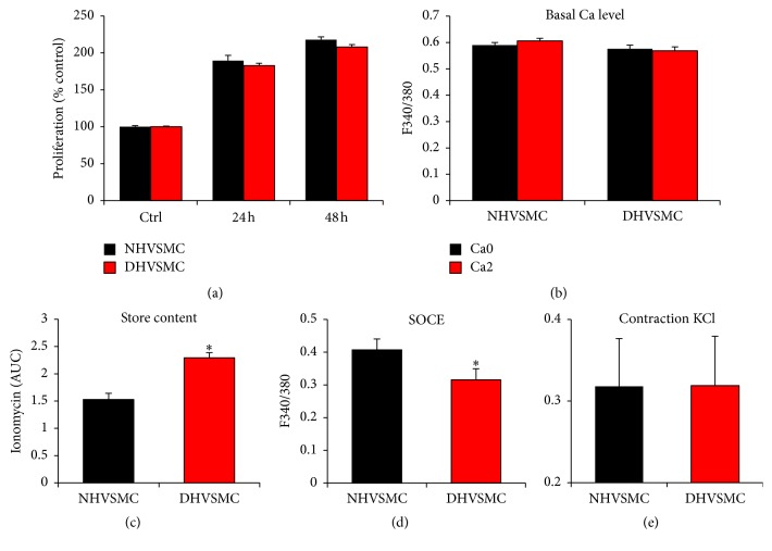
Figure 8.
Proliferation and Ca2+ signaling in VSMC cells from normal (NHVSMC) and diabetic (DHVSMC) human aorta. For all the tests human normal and diabetic VSM cells were cultured under NG as recommended by the supplier. Cell proliferation was determined 24 and 48 h after plating, using the WST-1 assay (a). For Ca2+ basal levels and Ca2+ store content, cells were loaded with 2 μM Fura-2AM for 30 min and the extent of Ca2+ release was determined following treatment of the cells with 2 μM ionomycin (b-c). For SOCE measurement, cells were loaded with 2 μM Fura-2AM for 30 min and SOCE was determined after store depletion with 1 μM thapsigargin (TG) (d). Cell contractility in response to 20 mM KCl was determined as described in Materials and Methods (e). Data are presented as mean ± SE from at least three independent experiments done each in triplicate. ∗p < 0.05.

