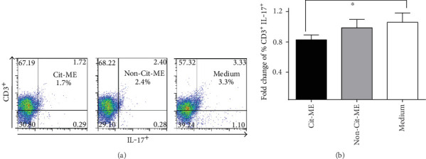
Figure 5.

Cit-ME downregulated Th17 cells in vitro. (a) Representative plots of CD3 and IL-17 staining. Positive staining is presented in the right upper quadrant of each plot with the percentage indicated. (b) Fold change of % CD3+ IL-17+ T cells (n = 11). Data are presented as mean values (∗p = 0.05).
