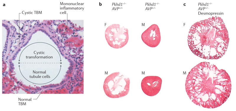
Figure 2. Features of early cyst formation.
a | Haematoxylin and eosin stain of a nascent renal cyst from a Han rat: a model of autosomal dominant polycystic kidney disease (ADPKD) caused by a missense mutation in ANKS6. The cells in the lower half of the image appear normal with brush borders typical of proximal tubules, whereas in the cystic upper half of the image the cells are flattened, with no brush borders and cramped nuclei. The extracellular matrix beneath the cystic cells is expanded and the tubule basement membrane (TBM) is thickened. Mononuclear inflammatory cells have invaded the interstitium adjacent to the cystic portion of the tubule. On microscopic analysis, a similar association between epithelial cell dedifferentiation and an abnormal underlying basement membrane has been observed in PKD1 and PKD2 murine models of ADPKD (J.J. Grantham & V.E. Torres, unpublished work). b | The first column (Pkhd1-/- AVP+/+) shows cross sections of kidneys from female (F) and male (M), 12-week-old rats, demonstrating an abundance of medullary cysts in the absence of Pkhd1 when arginine vasopressin (AVP) signalling is intact. In Pkhd1-/- AVP-/- rats (second column), which lack Pkhd1 and AVP signalling, very few cysts can be discerned. c | In Pkhd1-/- AVP-/- rats, administration of the AVP V2-receptor agonist, desmopressin, from 12–20 weeks of age results in the formation of visible cysts. Images reproduced with permission from Elsevier Ltd © Shizuko Nagao et al. Kidney Int. 63, 427–437 (2003).

