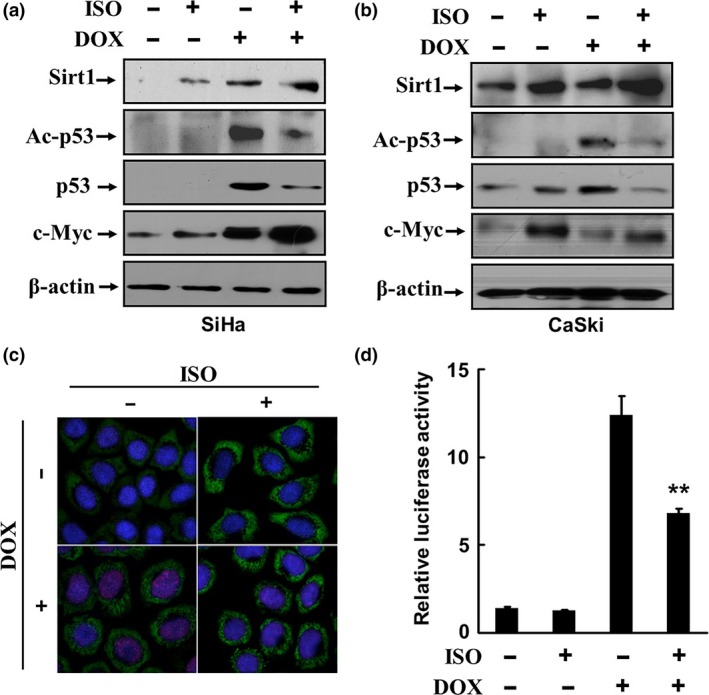
Figure 3.

Catecholamines inhibit DOX‐induced p53 acetylation and transcription‐activation activities. SiHa and CaSki cells were treated with 0.5 μM DOX for 18 h and then with 2.5 μM ISO for 6 h. The expressions of Sirt1, c‐Myc, p53, and acetylated p53 were analyzed by Western blot (a, b). The expression of Sirt1 (green) and acetylation of p53 (red) in SiHa cells was detected by indirect immunofluorescence staining and cofocal microscopy (c). (d) SiHa cells were transfected with a luciferase reporter plasmid containing consensus p53 DNA binding sites and thymidine kinase promoter‐Renilla luciferase reporter plasmid. The transfected cells were treated with 0.5 μM DOX for 18 h and then stimulated with 2.5 μM ISO for 6 h. The activities of p53 were determined by luciferase assays. **P < 0.01, compared to the cells treated with DOX alone.
