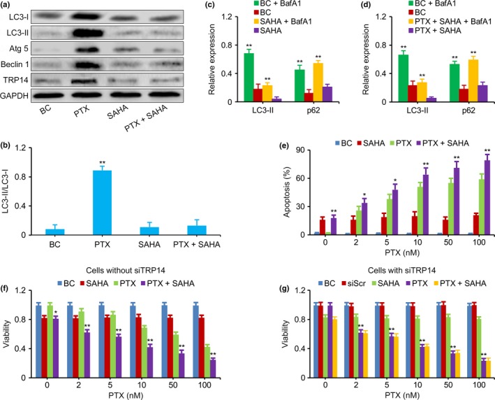
Figure 4.

Inhibition of TRP14 increased the antitumor effects of paclitaxel (PTX). (a,b) Western blot showed that the expression of LC3‐I, LC3‐II, Atg 5, Beclin 1, TRP14 and LC3‐II‐to‐LC3‐I ratio was not increased in SK‐N‐SH cells exposed to SAHA alone or SAHA combined with PTX; this is the opposite result to that obtained in cells exposed to PTX alone. GAPDH was used as the loading control. (c,d) LC3‐II and p62 in QDDQ‐NM cells measured by western blot. The increased levels of LC3‐II and p62 expression that resulted from BafA1 treatment were similar in cells exposed to SAHA (with or without PTX) compared with cells that were not exposed to SAHA (with or without PTX). (e) The apoptosis was enhanced in QDDQ‐NM cells exposed to PTX plus SAHA compared to PTX alone. (f) MTT assay showed that the viability of SK‐N‐SH cells treated with PTX combined with SAHA was lower than that of cells treated with PTX or SAHA alone at the doses of 2, 5, 10, 50 and 100 nM. (g) MTT assay showed that the viability of siTRP14‐treated QDDQ‐NM cells that were exposed to PTX alone at each PTX dose level was similar to that of cells exposed to PTX plus SAHA. BC, blank control, neuroblastoma (NB) cells without PTX and SAHA exposure. The results are presented as the mean ± SEM of three independent experiments. *P < 0.01, **P < 0.01, two‐tailed t test.
