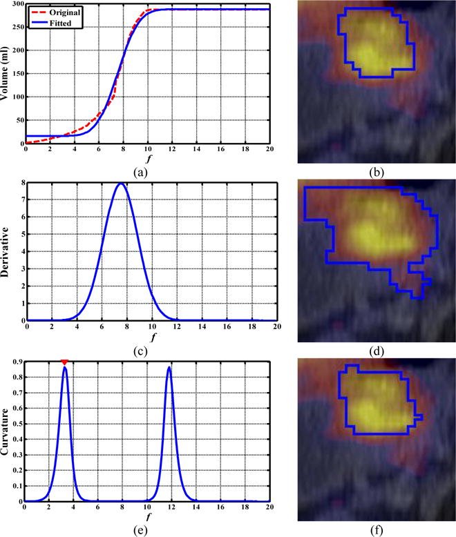
FIG. 10.

Comparison of the ORF obtained by ARG_MC and ARG_MD for a patient with esophageal cancer. (a, c, e): the f-volume curve (red line) and the fitted f-volume curve (blue line), its derivative, and its Menger curvature, respectively; (b, d, f) axial PET/CT view: manual contour, segmentation results obtained by the ARG_MD method (DSI=0.38) and ARG_MC method (DSI=0.89), respectively.
