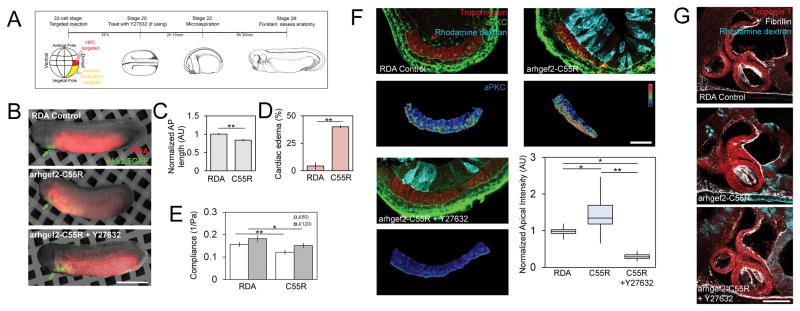
Figure 5. Targeted injections to perturb microenvironmental stress via actomyosin contractility accelerate MET.
(A) Experiment schematic: 32-cell stage injected to target either HPCs (red) or the anterior endoderm (yellow). (B) Transgenic nkx2.5-GFP embryos co-injected with endoderm targeted arhgef-C55R mRNA and rhodamine dextran (scale bar, 1 mm). Cardiac differentiation proceeds but arhgef2-C55R reduces AP length (C) and increased rates of pericardial edemas per clutch (D; N = 23 control, 35 arhgef2-C55R, 3 clutches). (E) Compliance of arhgef-C55R reduced (N = 8–9 embryos over 2 clutches). (F) Upper: tropomyosin, aPKC, and rhodamine dextran. Lower: tropomyosin-masked aPKC expression in psuedocolor LUT (scale bar = 50 μm). Apical aPKC increased in arhgef2-C55R, while Y27632 treatment can reverse effect (N = 7–11 embryos over 3 clutches). (G) Representative confocal sections of stage 39 tadpole heart morphology (scale bar = 100 μm). Error bars, ± SEM. See also Figures S1, S3B, S4 and S5.

