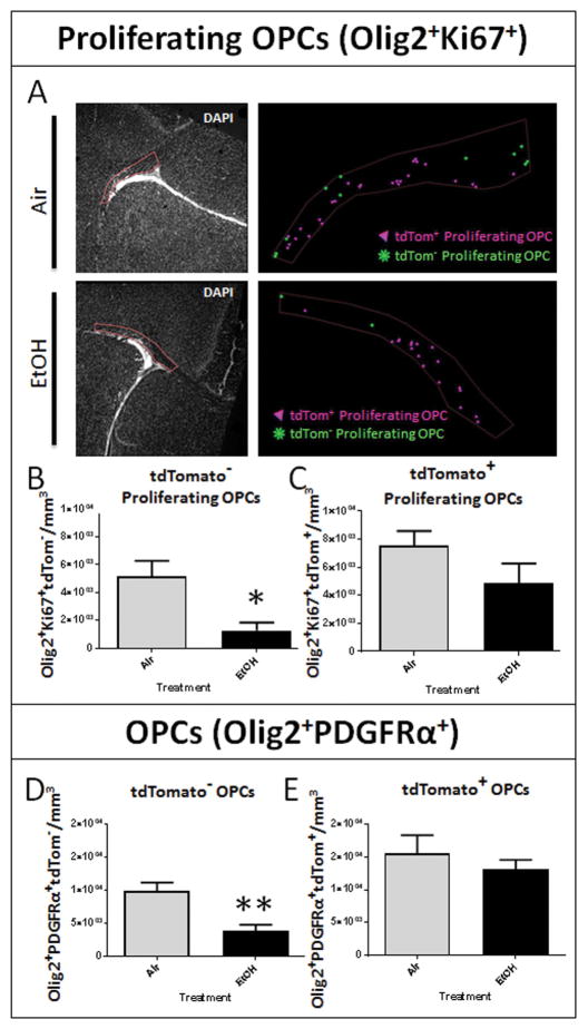
Figure 4.
Developmental EtOH exposure results in an acute and origin dependent loss of oligodendrocyte progenitor cells. (A) ROIs encompassing septal corpus callosum pertaining to the adjacent marker maps illustrating the distribution of tdTom+ and tdTom− proliferating OPCs (Olig2+/Ki67+) in air and EtOH exposed animals. (B) At P16, stereological analysis of immunohistochemically processed sections containing the corpus callosum at the level of the septal nuclei revealed a significant reduction in the density of tdTom− proliferating OPCs in the corpus callosum following EtOH exposure (*p = 0.0130), (C) whereas the density of tdTom+ proliferating OPCs was unaffected (p = 0.1696; n = 4–7 animals per treatment group). (D) At P16, EtOH exposure diminished the density of the total tdTom− OPC (Olig2+ PDGFRα+) population (**p = 0.0042; n = 4–6 animals per treatment group). (E) No discrepancy in tdTom+ OPCs between Air and EtOH exposed animals was observed (p = 0.4825; n = 4–6 animals per treatment group).

