
Figure 7. Model of the ABL-IRE1α axis.
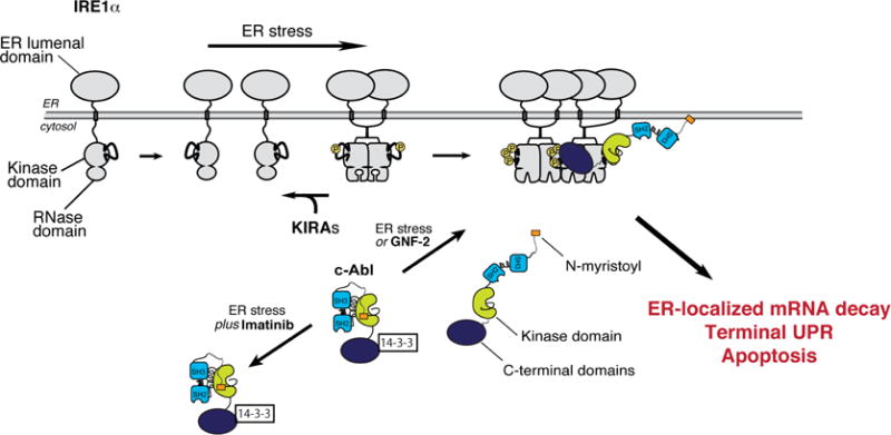
Under ER stress, c-Abl, dissociates from cytosolic 14-3-3 proteins to co-localize with IRE1α at the ER membrane, thus driving high-order oligomerization, ER-localized mRNA decay, T-UPR induction, and apoptosis. Imatinib prevents c-Abl’s re-localization to blunt the T-UPR. GNF-2 forces c-Abl localization to IRE1α, without ER stress, activating the T-UPR. Under ER stress or GNF-2, KIRAs block IRE1α hyperactivation and the T-UPR.