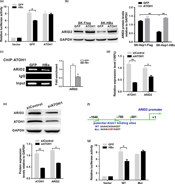
Figure 5.

HBx inhibited ARID2 promoter activity via an ATOH1‐dependent pathway. (a) Luciferase activity of the human ARID2 promoter reporter pGL3‐ARID2 in Huh7 cells. Huh7 cells were transfected with pGL3‐ARID2 and then co‐infected with AdGFP control or AdHBx together with AdATOH1. Luciferase activities were measured at 24 h after infection (n = 3, *P < 0.05). (b) ARID2 protein expression was determined by western blotting in Sk‐Hep1/SK‐Hep1‐HBx cells infected with AdATOH1 or AdGFP control (n = 3, **P < 0.01). (c) ChIP assays of cell extracts from Huh7 cells infected with AdHBx or AdGFP using anti‐ATOH1 antibodies. IgG served as a negative control. The relative fold enrichment (bound/input) was measured by qPCR. Data represent the means ± SDs (n = 3, *P < 0.05). (d) and (e) The mRNA and protein expression levels of ARID2 in ATOH1‐depleted SK‐Hep1 cells. SK‐Hep1 cells were infected with lentiviruses carrying ATOH1 shRNA or control shRNA. Cells were performed qRT‐PCR and Western blot assay (n = 3, *P < 0.05; **P < 0.01). (f) Schematic representation of the ARID2 promoter region with the potential ATOH1 binding sites indicated. (g) Luciferase assay of ARID2 promoter constructs with the wild‐type (WT) or mutated ATOH1 binding site in AdGFP‐ or AdHBx‐infected Huh7 cells. Data are shown as means ± SDs (n = 3 independent experiments; *P < 0.05 by Student's t‐test).
