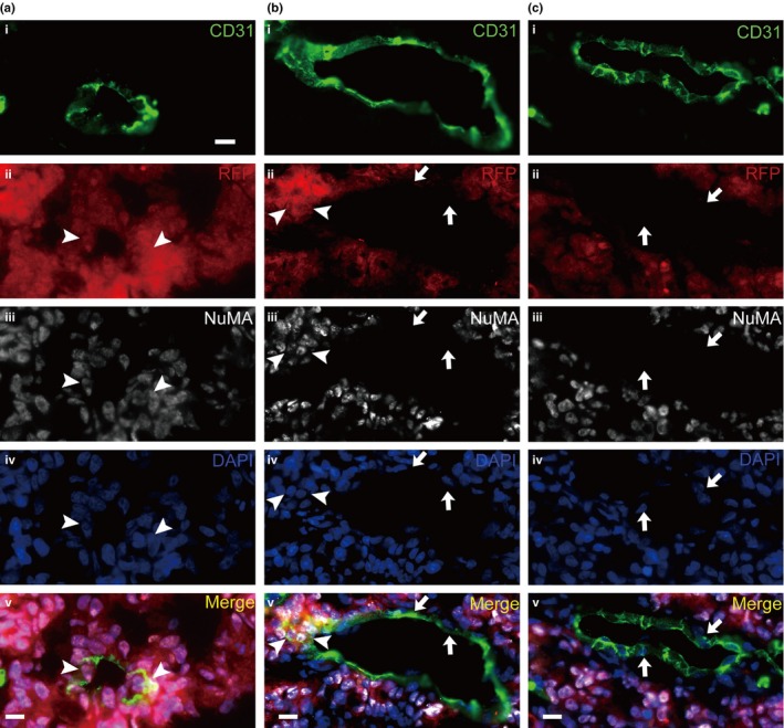
Figure 2.

Red fluorescent protein (RFP)‐labeled cancer stem cells of human colorectal carcinomas (CoCSC) generate RFP+ endothelial cells (EC) in xenograft tumors. Representative immunofluorescence micrographs of tumor xenograft sections are shown. The EC expressed CD31 (a–c(i)) and the cells originating from CoCSC‐expressed RFP (a–c(ii)) and NuMA (a–c(iii)). DAPI was used as the nuclear marker (a–c(iv)). The images were incorporated (a–c(v)). Arrowheads indicate NuMA +/RFP + EC and arrows indicate NuMA −/RFP − ECs. Scale bars: 20 μm.
