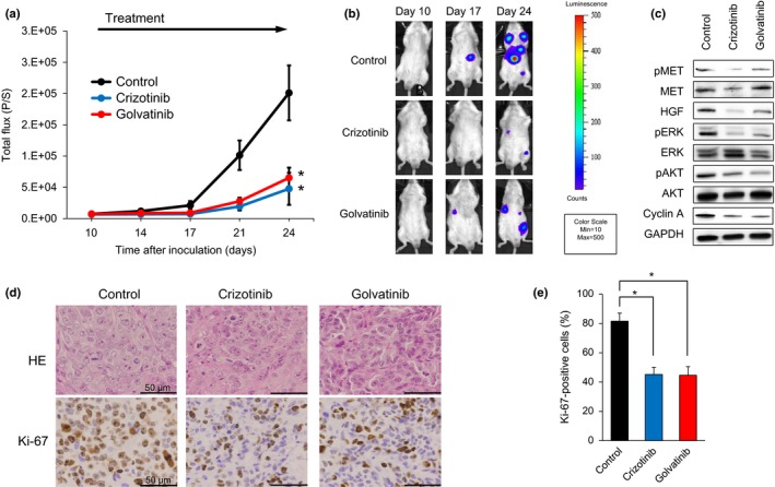
Figure 5.

MET inhibitors significantly inhibit tumor progression of SBC‐5 small‐cell lung cancer cells in an in vivo multi‐organ metastasis mouse model. (a) EGFP‐Eluc‐transfected SBC‐5 (SBC‐5/EGFP‐Eluc) cells were injected i.v. into natural killer cell‐depleted SCID mice. At 10 days after inoculation, the mice were randomized into vehicle (control), crizotinib (100 mg/kg), or golvatinib (50 mg/kg) treatment groups (n = 6 per group), and began treatment once daily by oral gavage. Luminescence was evaluated twice weekly. Bars indicate standard error. *P < 0.05 by Mann–Whitney test, versus control group. (b) Representative images of mice showing merged bioluminescence and photograph. (c) Liver tumors were resected from mice 3–4 h after treatment on day 35. Relative levels of proteins observed in each tumor were determined by Western blot analysis. AKT, protein kinase B; HGF, hepatocyte growth factor; p, phosphorylated. (d) Representative images of liver tumors immunohistochemically stained with antibodies to human Ki‐67. Bar = 50 μm. (e) Quantification of proliferating cells in liver tumors, determined as the observed percentage of Ki‐67‐positive cells. The data shown represent the mean of five areas ± SD. *P < 0.05 by Student's t‐test, versus control group.
