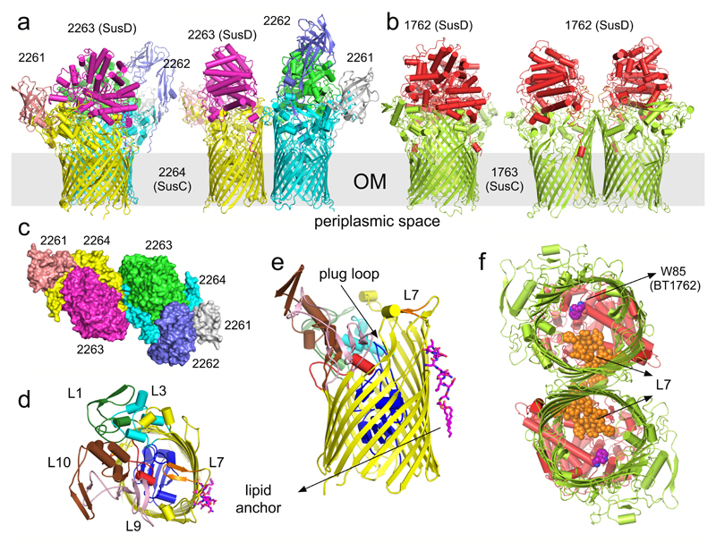
Figure 1. Overall architecture of SusCD complexes.
a, b, Cartoon views from the side for BT2261-64 (a) and BT1762-63 (b) rotated by 90º, with individual subunits labelled. OM, outer membrane. c, Surface representation of BT2261-64 from the outside of the cell. d, e, Architecture of BT2264 (SusC-like) viewed from the extracellular side (d) and from the plane of the membrane (e) with several extracellular loops indicated (L). The plug domain in the 22-stranded β-barrel is dark blue, and the N-terminal ten residues of BT2263 (SusD-like) including the lipid anchor are shown as stick models in magenta. Loop L7 is shown in orange. f, View of the BT1762-63 dimer from the periplasmic side. The L7 loops (orange) of BT1763 (SusC-like) and the functionally important residue Trp85 in BT1762 (SusD-like) are shown as space-filling models.

