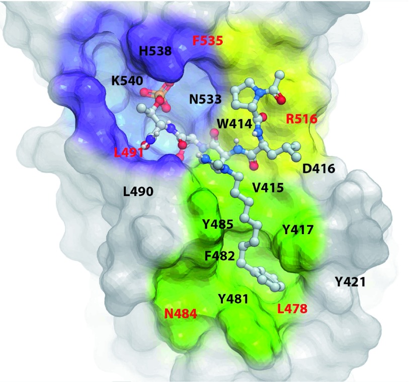
Figure 5. The structural model of polo-box domain (PBD) in complex with 4j.
The X-ray cocrystal structure of the PBD + 4j complex (PDB: 3RQ7) shows a “Y-shaped” binding pocket composed of three discrete but interlinked binding modules—namely, a p-Thr/p-Ser-binding module (violet), a Pro-binding module (yellow), and a hydrophobic channel (green). Residues highlighted in red are specific to Plk1 PBD. Inhibitors designed to bind to more than one of the three binding modules could possess a superior binding specificity because of the specific requirement of the shape and geometrical arrangement of their binding moieties (see text for details).

