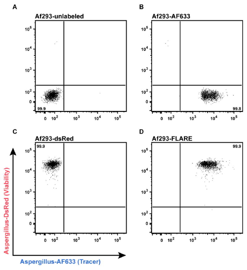
Figure 1. Labeling efficiency of FLARE conidia.

Flow plots depict fluorescence intensity of (A) unlabeled, (B) AF633-labeled, (C) DsRed-labeled, and (D) FLARE conidia in AF633 (X-axis) and DsRed (Y-axis) channels using a BD LSR II flow cytometer equipped with 532 nm and 633 nm lasers.
