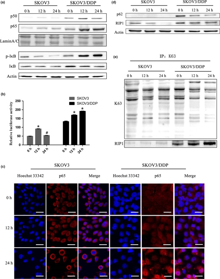
Figure 1.

Cisplatin treatment activates the NF‐κB pathway and an increase in RIP1K63‐linked ubiquitination in SKOV3/DDP cells. (a) Cells were treated with cisplatin (6 μg/mL) and expression of NF‐κB p65 and NF‐κB p50 in the nucleus and IκB, p‐IκB in whole cell lysates was analyzed by western blotting. (b) SKOV3 and SKOV3/DDP cells were transiently transfected with a pNF‐κB‐Luc vector. The luciferase activity was assessed and normalized on the basis of Renilla luciferase activity (mean ± SD, n = 3. *P < 0.05, vs control). (c) SKOV3 and SKOV3/DDP cells were stained with Hoechst 33342 and antibodies against p65. They were observed under confocal laser microscopy (scale bar, 25 μm). (d) SKOV3 and SKOV3/DDP cells were immunoblotted (IB) with anti‐p62 and anti‐RIP1. (e) Immunoprecipitation (IP) was performed using the anti‐K63 antibody followed by western blotting using anti‐RIP1 antibody.
