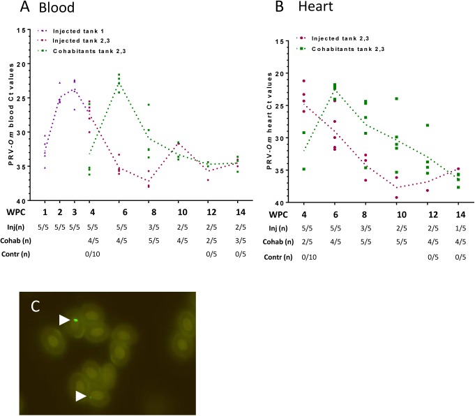
Fig 2. Rainbow trout long-term study (trial 2): PRV-Om in blood and heart.
A) Virus analysis performed by RT-qPCR targeting PRV-Om segment S1 in blood from virus-injected fish from three tanks (Tank 1: purple, parallel tanks 2 and 3: red) and cohabitants from tanks 2 and 3 (green). Large dots indicate Ct values of individual fish and dotted trend lines the mean Ct value of virus-positive fish. The table below shows number of positive fish per sampling point. B) PRV-Om Ct values in heart of fish from tanks 2 and 3. C) Detection of PRV-Om in rainbow trout red blood cells using rabbit antiserum targeting PRV-Ss σ1. Arrows mark positive staining.

