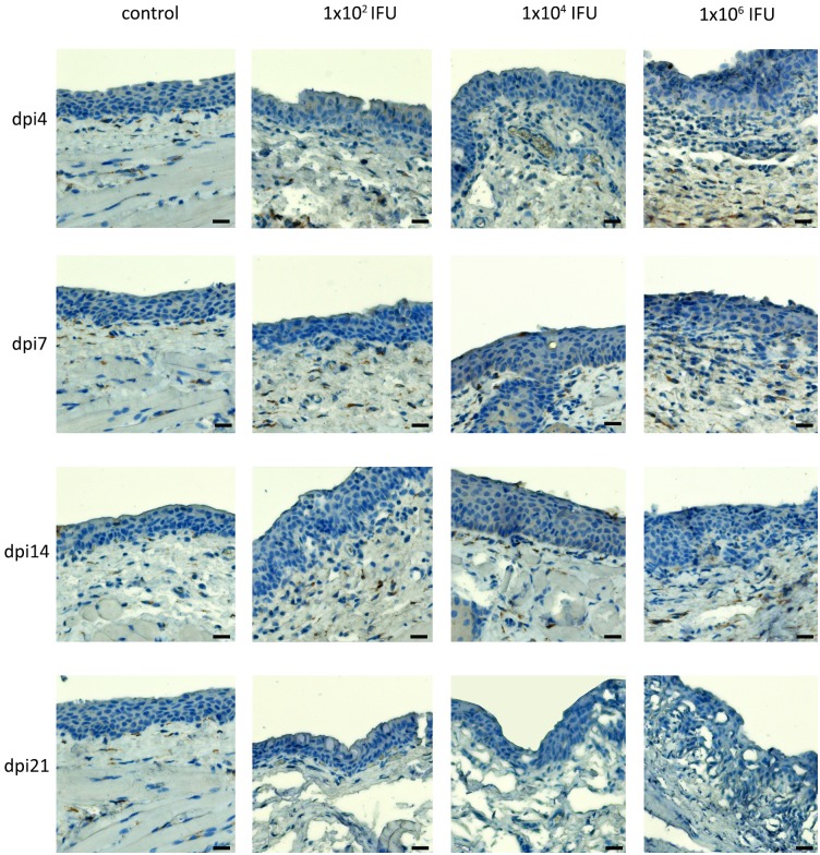
Fig 3. The abundance of neutrophils in the conjunctiva and CALT of guinea pigs infected with a single ocular instillation of three different C. caviae doses.
The neutrophils infiltration was evaluated in paraffin-embedded conjunctival sections by immunohistochemical staining for myeloperoxidase, a neutrophil-specific enzyme. Conjunctival sections were prepared from samples collected at screening time points within the post-infection period (day 4 –dpi4, day 7 –dpi7, day 14 –dpi14, day 21 –dpi21). ExtrAvidin®−Peroxidase/DAB system was used for visualisation of myeloperoxidase presence.

