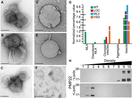
Fig. 1. PMP22 forms ordered assemblies upon reconstitution into lipid vesicles.

(A to C) Examples of protein-lipid MLAs created when PMP22 is reconstituted into 4:1 POPC/ESM vesicles via the dialysis method and visualized by negative stain EM. (D) Representative image of multilamellar vesicles (MLVs) prepared in the absence of protein via the dialysis method [lipid-only control (LOC)]. (E) MLVs prepared by spontaneous bilayer formation through hydration of lipids with water. (F) Control assemblies containing 4:1 POPC/ESM and the tetraspan VSD of KCNQ1 reconstituted via the dialysis method. Scale bars (all panels), 100 nm. (G) Quantification of the relative percentage of MLAs present in a series of negative stain EM images of wild-type (WT) PMP22, LOC, MLVs, and the tetraspan VSD domain of KCNQ1. All individual object counts were converted to percentage of total counts for a particular sample and were normalized to the percentage of total counts represented by MLAs in the WT PMP22 control, which was set to 1.0. Green, WT; red, LOC; blue, MLV; orange, VSD. (H) Sucrose gradient analysis of PMP22 reconstituted for 10 days without (−; top) or with (+; bottom) lipids. Fractions were collected from top (low density) to bottom (high density) and analyzed by SDS–polyacrylamide gel electrophoresis (PAGE) with silver staining.
