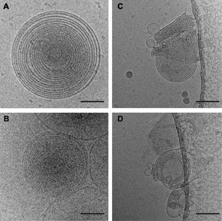
Fig. 2. Differences between MLVs and MLAs are visible by cryo-EM.

(A and B) Representative images of vitrified MLVs prepared in the absence of protein via the dialysis method (A) or by spontaneous bilayer formation through hydration of lipids only with water (B). (C and D) Examples of MLAs created when PMP22 is reconstituted into 4:1 POPC/ESM vesicles via the dialysis method and visualized using cryo-EM. Scale bars (all panels), 100 nm.
