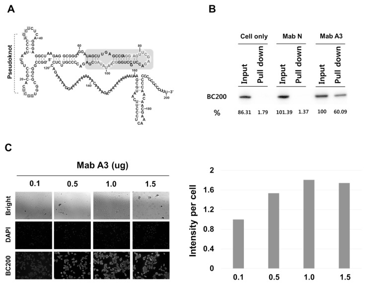
Fig. 1.
Specific recognition of BC200 RNA by the antibody, MabBC200-A3, in HeLa cells. (A) Possible secondary structures of BC200 RNA. The blue- shaded region is the domain, recognized by the antibody MabBC200-A3. Protected regions by the MabBC200-A3 antibody are highlighted in red letters. (B) HeLa cell lysates were immunoprecipitated with MabBC200-A3. RNAs were purified from the immunoprecipitates and subjected to Northern blot analysis. Cell only, without antibody. Mab N, a negative control antibody. Mab A3, MabBC200-A3. (C) Cells treated with increasing amounts of MabBC200-A3 were incubated with Cy™2 AffiniPure Donkey Anti-Human IgG and subjected to confocal microscopy. BC200 RNA is represented by green fluorescence. DAPI was used for nuclei staining.

