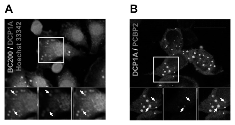
Fig. 4.
Co-localization between BC200 RNA and DCP1A, and between hnRNP E2 and DCP1A. (A) HeLa cells were transfected with a DCP1A-mCherry-expressing construct, treated with MabBC200-A3, and incubated with Cy™2 AffiniPure Donkey Anti-Human IgG. The nuclei were stained with Hoechst 33342. BC200 RNA, DCP1A-mCherry, and the nuclei were visualized as green, red, and blue fluorescence, respectively, under confocal microscopy. Co-localization is shown by arrows. (B) HeLa cells were transfected with hnRNP E2-eCFP- and DCP1A-eGFP-expressing constructs, and the cells were subjected to confocal microscopic analysis. hnRNP E2-eCFP and DCP1A-eGFP were visualized as blue and green fluorescence, respectively. Co-localization is shown by arrows.

