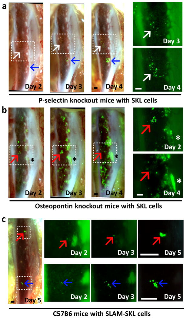
Figure 7. SKL cell engraftment in abnormal HSC niches.
(a) Time-lapse in vivo imaging of SKL cell engraftment in P-selectin knockout mice (n=5). Cells engrafted in the central marrow area engrafted and proliferated much slower (white arrow) than a cell that homed to the endosteal region (blue arrow). Higher magnification pictures of the boxed area on the right side show aberrant engraftment in the central marrow region (white arrow). 6×10⁴ GFP+ SKL cells were injected at Day 0 after lethal irradiation. (b) In vivo imaging of SKL cell engraftment in OPN knockout mice showing rapid proliferation of SKL cells (n=4). Higher magnification pictures of the boxed area on the right side show clusters of SKL cells at the central marrow region disappearing at day 3 (red arrow). SKL cells in the endosteal regions showed rapid proliferation (asterisk). 3×10⁴ GFP+ SKL cells were injected at Day 0 after lethal irradiation. (c) 3×10³ GFP+ SLAM-SKL cells were injected at Day 0. Engraftment and proliferation of individual SLAM-SKL cells (red/blue arrows) were observed till Day 5.

