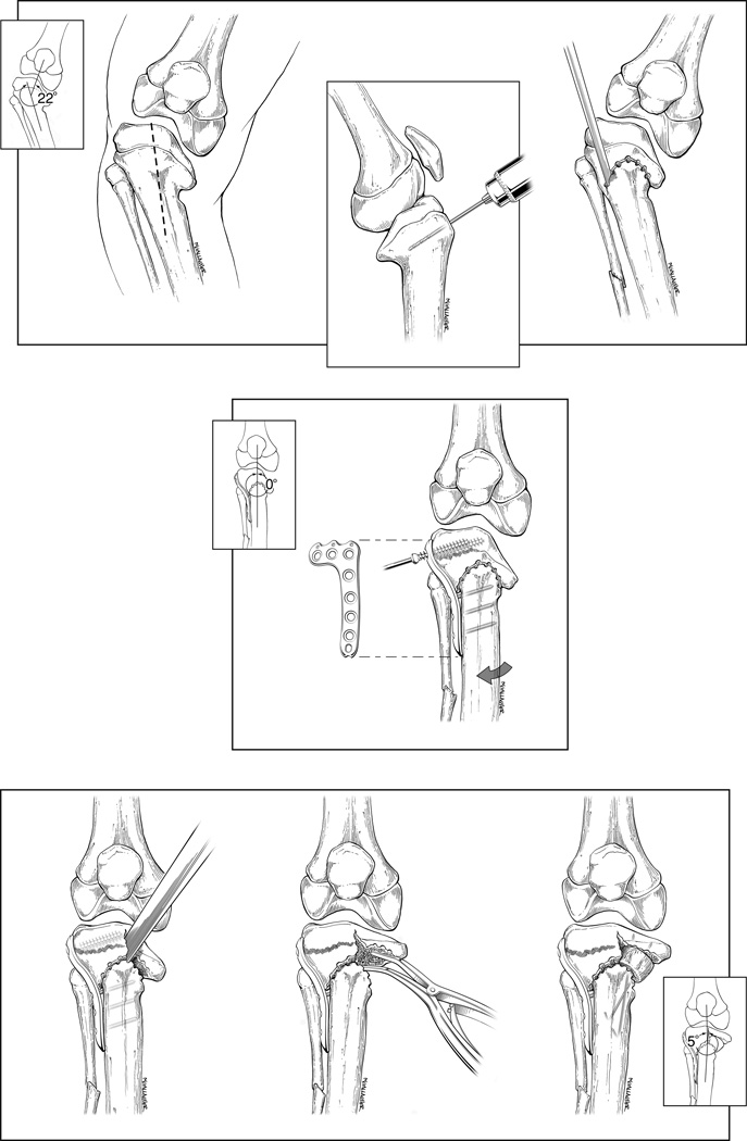
Fig. 2.
A–G Double tibial and fibular osteotomies surgical steps. (A) Skin incision. (B) drill bit is placed at apex of planned dome osteotomy and parallel to joint surface. (C) Dome osteotomy is outlined by drill holes and completed by osteotome. (D) lateral tibial plateau plate is applied with leg in corrected position and proximal to the growth plate. (E) Vertical osteotomy is performed from the apex of the dome osteotomy to subchondral bone. (F) Spinal laminar spreader is used to fully elevate the medial tibial plateau and corrects the posterior procurvatum. (G) elevated medial tibial plateau is supposed with bone graft-materials and stabilized with Kirschner wire(s).

