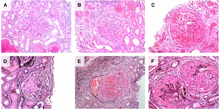
Figure 3.
Renal histopathology in anti–glomerular basement membrane (anti-GBM) GN. (A–C) Hematoxylin and eosin–stained sections demonstrating (A) segmental fibrinoid necrotizing lesion in early anti-GBM GN; (B) small, circumscribed cellular crescent; and (C) large, circumferential cellular crescent. (D–E) Demonstrate the use of Jones methylamine silver stain to delineate glomerular and tubular basement membranes, clearly identifying a segmental area of extracapillary proliferation (D). (E) Demonstrates obliteration of the glomerular architecture and rupture of Bowman’s capsule, with extravasation of red blood cells into the urinary space, and significant peri-glomerular inflammation. (F) Adjacent glomeruli with synchronous cellular crescent formation typical of anti-GBM disease.

