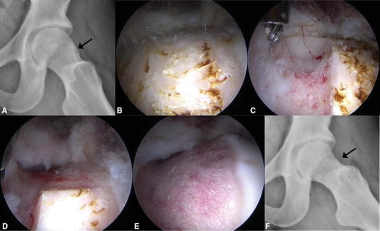
Fig. 1A–F.

A female patient with cam FAI was treated with femoroplasty using the sclerotic subchondral cortical bone thickness as a guide for resection depth. (A) Her preoperative frog-leg lateral radiograph shows the sclerotic region of the cam lesion (arrow). Intraoperative arthroscopic views from a 70° scope through the anterolateral portal show (B) the sclerotic region, (C) the initial resection trough where the depth was based on the sclerotic subchondral cortical bone thickness, (D) continuation of the resection based on trough depth and patient anatomy, and (E) the completed resection. (F) The patient’s postoperative frog-leg lateral radiograph shows where the sclerotic region has been removed (arrow).
