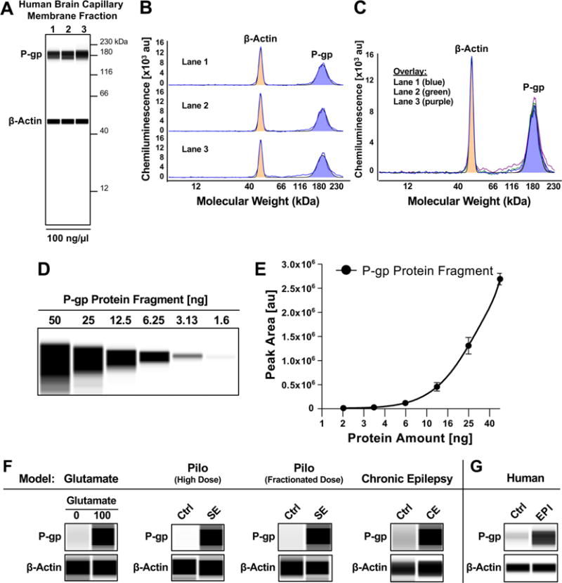
Figure 5.

P-gp expression and activity in brain capillaries in animal models and human epilepsy. (A) Wes images showing P-gp and β-actin protein expression in isolated human brain capillaries. (B) Electropherogram for each lane and C) overlay of lanes 1–3. (D) Concentration series and (E) standard curve for human P-gp protein fragment used for quantitative analyses. (F) Wes images showing upregulation of P-gp protein expression levels in all four models: (1) ex vivo glutamate model, (2) high-dose SE model, (3) fractionated SE model, and (4) CE model. (G) Increased P-gp protein expression in brain capillaries isolated post-mortem from brains of individuals that experienced generalized seizures compared to seizure-free control individuals. β-actin was used as protein loading control.
