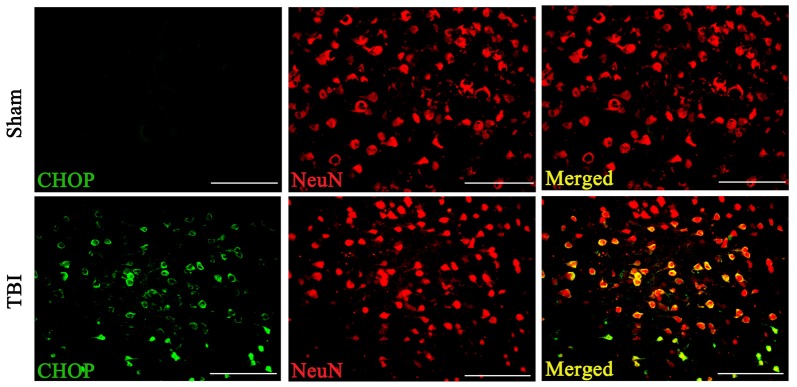
Figure 3.
Coronal brain sections from C57/BL6 mice (Bregma −2.20) were double immune stained for CHOP protein (green) and NeuN (red). Levels of CHOP in neuronal cells were assessed immunofluorescence. Images show CHOP positive neuronal cells was considerably higher in the lesion area of the cerebral cortex 72 h following TBI. n = 5 for each group. Scale bar = 50 μm.

