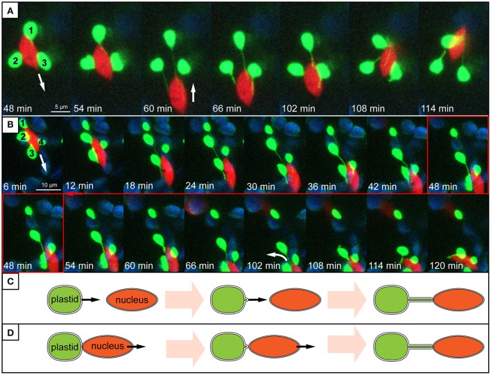
Figure 8.
Stromule behavior seems linked to nucleus movement. (A,B) Images are maximum intensity projections along the z-axis, taken from a time series of z-stacks obtained by a confocal laser-scanning microscope. Bright green plastids and red nuclei are organelles of the upper epidermis of an A. thaliana wild-type plant transformed with the pLSU4::pn plasmid (blue channel, chlorophyll fluorescence; red channel, H2B:mCherry fluorescence; green channel, FNRtp:eGFP fluorescence). The white arrows indicate the direction of nuclear movement. Larger, round, blue signals represent underlying mesophyll plastids, where chlorophyll signal is brighter than the FNRtp:eGFP fluorescence signal. (A) Series of images taken from Supplementary Movie 2. It demonstrates how nucleus movement “pulls” stromules out of a group of stationary plastids (1–3) and how stromules shorten, when the nucleus changes its direction. (B) Series of images from Supplementary Movie 3 showing how a nucleus moves away from a group of plastids (1–4). Plastids 2–4 keep their extended stromules and follow the moving nucleus. Plastid 1 stays stationary and looses its stromule after the nucleus moved more than 12 μm away from it. (C,D) Schematic representation of two possible ways how nucleus associated stromules might be formed. (C) Scenario 1: stromules are formed by plastids residing further away from the nucleus growing toward (“reaching out”) and connecting to the nucleus. (D) Scenario 2: nucleus associated plastids are anchored to the surface of the nucleus and due to nucleus movement a stromule is pulled out of the surface of the stationary plastid.

