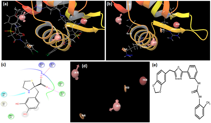
Figure 6.

Energy Based Pharmacophore development. (a) Multiple conformations of I-20 (IC50-1 µg/ml) bound to the DNA binding surface of IdeR. (b) Various pharmacophore points in proximity with Ser 37, Pro 39, Gln 43, Arg 27 and Ala 28. (c) 2-D Interaction map of I-20 with various residues of the DNA binding surface of IdeR. (d) Final 5-Point pharmacophore taken for subsequent screening of the drug like database of ZINC. (A16 and A11 refers to Acceptor feature, R48 and R50 refers to Aromatic ring feature and N38 refers to negative ionizable group). (e) 2-D structure of IA-8 3-[3-[5-(1, 3-benzodioxol-5-ylmethyl)-1, 3, 4-oxadiazol-2-yl]phenyl]-1-(o-tolyl) urea with an IC50 of 60 µg/ml.
