FIGURE 2.

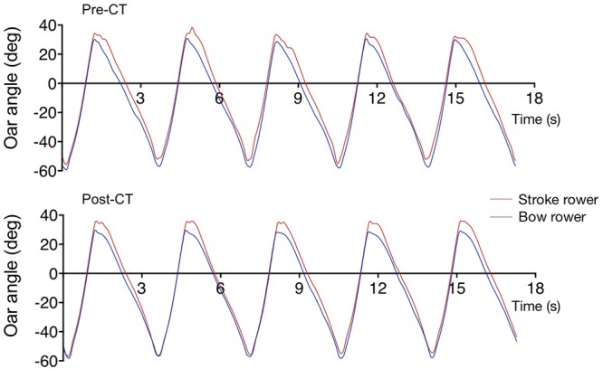
Samples of oar movement of the stroke (red) and bow (blue) rowers for five subsequent strokes during the pre-CT (Upper) and post-CT (Lower) sessions.

Samples of oar movement of the stroke (red) and bow (blue) rowers for five subsequent strokes during the pre-CT (Upper) and post-CT (Lower) sessions.