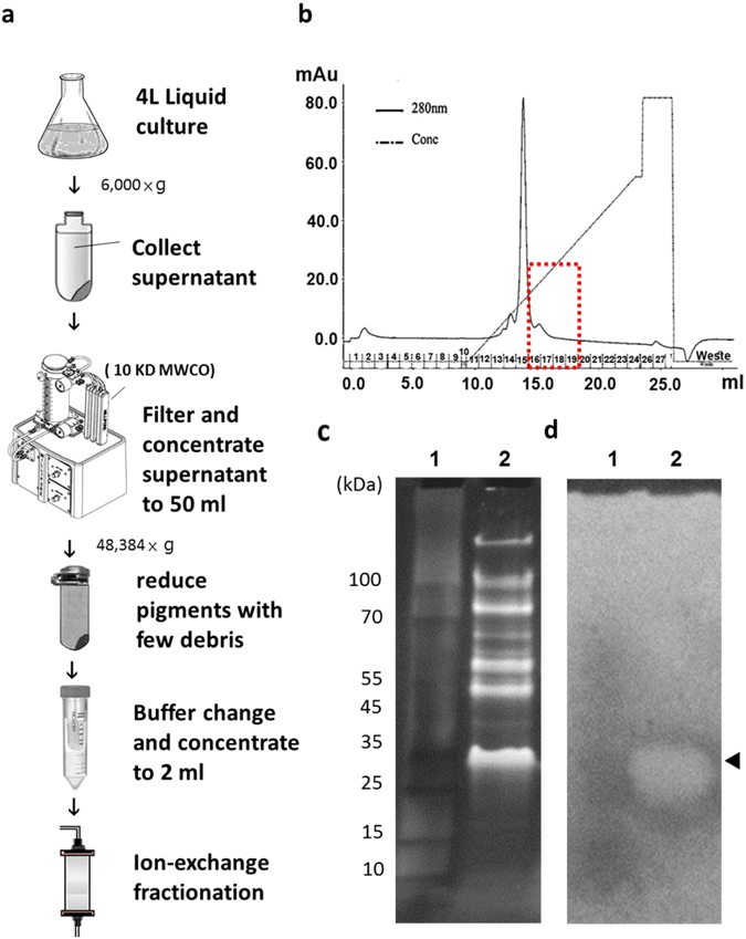
Figure 2.

Purification of keratinase from culture medium and keratinase identification. (a) An overview of keratinase purification from the overnight culture medium. Each step is described specifically in Materials and Methods. (b) Elution profile of MtaKer after chromatography using S-Sepharose column. The proteins were eluted with a linear salt gradient. The fraction No. 15 (the major peak) is the contaminating carotenoid pigment that was eluted by 200 mM NaCl. The red box indicates the collected peak (fraction No. 16–19) containing keratinase activates. (c) The proteins from the collected peak were further separated by 4–20% gradient SDS-PAGE to determine the size or molecular weight of unknown proteins. Lane 1: protein markers; lane 2: the collected peak with keratinase activity. (d) The keratinolytic enzyme profile was evaluated by replica-agarose-zymogram analysis on the 1% agarose with keratin powder/casein as substrates. The keratinolytic activity can be visualized as a clear zone (arrowhead) and the corresponding band on SDS-PAGE was subjected to standard proteomic analysis to identify the amino acid sequence of MtaKer.
