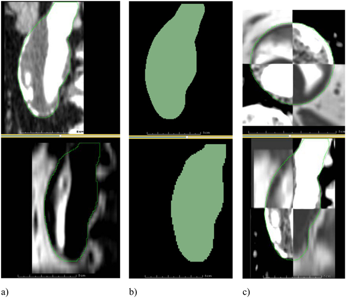
Figure 10.

MRI to CT registration. (a) A sagittal slice of an AAA in a contrast-enhanced CT image (top) and the corresponding slice in the MR image (bottom). The contours of the AAA segmentations are shown in green. The two images occupy different positions in space and need to be registered. (b) The segmentations of the CT (top) and MR (bottom) images. The two label maps are registered and the resulting transform is saved. (c) The transform obtained from the label map registration is used to register the MRI dataset to the CT dataset. A checkerboard display of the two images is used to verify the registration result in axial (top) and sagittal (bottom) views, with the contour of the AAA segmentation shown in green.
