Fig. 3.
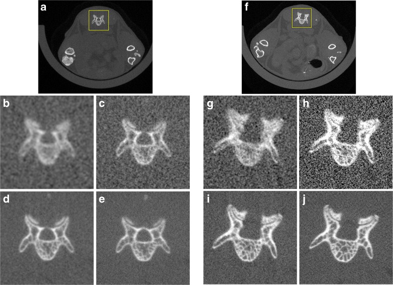
Example images of a transaxial rostral slice through L6 showing subvolume reconstructions at the highest resolution possible. a The low magnification was then cropped (yellow box) for display purposes to show the b 8-s scan, 36 μm isotropic voxel size, 10 mGy, c 18-s scan, 18 μm isotropic voxel size, 22.4 mGy, d 2-min scan, 18 μm isotropic voxel size, 130.4 mGy, or e 4-min scan, 9 μm isotropic voxel size, 269.1 mGy. f The high magnification was then cropped (box) for display purposes to show the g 8-s scan, 18 μm isotropic voxel size, 38.9 mGy, h 18 s scan, 9 μm isotropic voxel size, 87.1 mGy, i 2-min scan, 9 μm isotropic voxel size, 508.2 mGy, or j 4-min scan, 4.5 μm isotropic voxel size, 1048.2 mGy. All images are displayed with a level of 1000 HU and a window of 4000 HU with no filtering applied.

